The technique that produced the first female fetus *
Table of Contents
The technique that produced the first female fetus *
by Barbara Duden
(*) This article is a section of the chapter entitled “Schwangerschaft: die Gute Hoffnung und die Diagnose” from Barbara Duden Die Gene im Kopf – der Fotus im Bauch: Historisches zum Frauenkorper, Hannover, Offizin-Verl., 2002, pp.69-83. Jutta Mason was kind enough to translate it.
In rummaging through old atlases in search of how anatomists once viewed the unborn child, I ran into a surprisingly empty space. What is naturally “seen” today as an embryonic form and as the first stage of “the human being” lay in a blind spot for centuries: a being devoid of any human proportion — fat-headed, stumps for arms, bow-legged —was not recognized as a child in the making. The child-to-be was shown in many ways in anatomical treatises and atlases, but the pregnant woman's body was never the field/canvas/context for human developmental stages. Then, a Frankfurt anatomist, Samuel Thomas Soemmerring, brought the unrelated, motherless fetus into a new space of a-perspectival objectivity, using a new drawing technique that has no standpoint (Standpunklosen).
Two series of lectures in 1992, one at the University of Göttingen, the other at the Freie Universität Berlin on “Women's and Gender History” gave me the occasion to point out the radical change in the history of women and pregnant women that was effected by the technical production of the “fetus.” The woman's coming child, a matter of her heart’s desire, appears here in the unperceivable because constructed space of a-perspective objectivity.
The technique that produced the first fetus (1799)
It is probably ten years ago that I began to immerse myself ever more deeply into the history of the body in modern times. By then, women historians whom I admire had already declared this topic in women's history to be closed. “Do we really have to unpack the history of women through their bodies?” asked Arlette Farge in 1984. It was a rhetorical question for her, just before shelving the issue. Yet the historicity of the body experience captivated me, because for me it became the key to the gender relations that are typically reflected today in the conflict over abortion, in the discussion about reproductive medicine, or in the so-called “Erlanger baby” (when a pregnant mother is brain-dead but kept alive to bring the baby to term). As baby, the new being is a matter of the heart in relation to a woman, whereas the Fetus creates its niche as a problem of hormonal regulation.
I want to interpret the first depiction of a woman’s “fetus”. The figure comes from a work by Samuel Thomas Soemmerring.1) As far as I can see in the history of the unborn, it is the first such depiction of a female fetus. As you know, Eve went through no fetal stage, for she was made during sleep as – I quote from the medieval sources – collateralis et aequalis of man's flesh, and not of dust like man. According to the current state of historical research, the daughters of Eve, as we were called, went through no fetal stage up to 1799. I'm speaking ab mundo condito, since the creation of the world. Eve, according to the Jewish calendar, was created about 6000 BC., and over the generations from Sarah, to Anna, to Mary, the mother of Jesus, and for another 1799 years – a nearly eight-thousand-year history – there was no fetus. Of course, before Soemmerring, there were other depictions of women in utero: I am thinking of the little girl in a five-picture series from the 12th century, demonstrating “childhood” in an upside-down cupping glass;2) I am thinking of a baroque panel on which an Unborn baby with a slit belly proudly points her index finger at her hymen and other female innards. But neither the demure spinster in the miniature nor the little exhibitionist from Adrian Spieghel's 1626 treatise on the formed fetus do I call “fetuses”. And, of course, the kicking putto from a 1604 Paduan print isn't a fetus either; “c” shows the sweat in which it swims (sudor cui innatat fetus) and “b” what was then called the “belly root” in anatomical German, i.e., the umbilical cord.3) I classify all of these figures as symbolic representations of unborn children. I contrast them with the depiction of the pre-child, i.e., the fetal, the embryonic form, as it was first depicted by Soemmerring.
What I want to offer for discussion has two sides. First, the fascination of a German scholar in 1799 with sex determination ab ovo; second, the break in the history of gender relations, which becomes visible in the appearance of the fetal figure. This engraving of a female fetus is not only a new appearance as a fetus and as a prenatal woman, but also as a diagram. What is represented is not what the anatomist saw, but as I will show, what he measured. Based on this figure/image, I want to discuss the emergence of a singular paradox around 1800: technogenesis, the technologically determined emergence of the simulacrum of gender neutrality in intimate connection with the emphasis on gender difference.
To sketch out this novel construction of a new gender relationship, I want to proceed in three steps: I want first to present my source; secondly, I want to talk about the contrast between icons and constructs of the unborn in the history of anatomical graphics; thirdly, I want to say something about the method of image interpretation. I want to show that I can only get at this picture if I distinguish between an iconographic and an iconological analysis of anatomical graphics. Only then can I interpret the two panels by Samuel Thomas Soemmerring from which the figure of the little fetus comes.
The Sources: Soemmerring’s List
In 1799 Soemmerring published the Icones Embryonum Humanorum in the Elephantenfolio. At the age of 44, he was the leading anatomist in Germany, with whom Goethe had been in correspondence about the premaxillary bone since his visit in 1783. Soemmerring's theses involved important contemporaries in controversies: about the degeneration in the physique of a Moor living in Kassel Wilhelmshöhe, which Soemmering thought was the result of climate4); about the cultural deformation of ribs, waist and hips produced by cinching the waist; about the organ of the soul in the body; about eye and ear; about the possibility of electric telegraphy.
The Prefatio to the Icones, the foreword to the foundational work of the “Illustrations of Human Embryos' ' begins with a listing of its predecessors/antecedents. Soemmerring names 37 anatomical treatises in which human embryos or women's eggs were not only described, but also presented graphically. The list begins with Hieronymus Fabricius ab Aquapendente, a successor of Vesal on the chair of anatomy in Padua at the beginning of the 17th century, from whose De Formato Foetu comes the putto, which I will later use as a contrast to Soemmerring. Soemmerring also notes that he had seen the copies engraved in copper by Jan Wandelaar in 1738.
Soemmerring's chronological list of illustrations of eggs and embryos is still unsurpassed today. I have supplemented Soemmerring's list with further illustrations from the 15th to 17th centuries and created a complete corpus of graphic representations of the unborn in prints since 1492. During this investigation, I have come to the conclusion that Soemmerring is correct in claiming that he was the first to produce a series of figures showing the “human body from the third week to … the sixth month.”5)
Astigmatism, or the contrast between child and fetus
This claim to be the first, on the part of the professor, who was already well established through marriage in Frankfurt, is even more surprising because gynecological illustrations had long been in circulation in scholarly treatises and popular broadsheets. Beyond that, cheap and illustrated midwifery handbooks had also been available since the 17th century. These showed not only the “birth organs of every woman (illustrated), how such are internally formed and situated”, but also “how the child fits and rests in the womb.”6) And yet Soemmerring is right: nascituri, children getting ready to be born, were to be seen, but what was never seen was what he himself wanted to show, the embryo. The contents of the pregnant mother remained an emblem.
Thanks to the art of dissection and preparation, thanks to the rules of perspective and cross-hatching, thanks to the increase in plasticity and tactile quality in the transition from woodcut to copperplate engraving, it was precisely during this period that ever more realistic depictions of entrails and the most delicate tissues emerged. The vesicles on the surface of epithelia and the capillaries in the transparent membrane of the uterus had been pricked out with astonishing fidelity since the end of the 17th century - mostly based on dissection. And yet, over a period of 300 years, despite the availability of these graphic means, the figure that has settled as a fact in the minds of pregnant women in 19th-century textbooks and is today mediated onscreen, remained unseeable.
Up until Soemmerring, the graphics showed the child to come. The child was not depicted but appeared symbolically, emblematically, fantastically, and occasionally, in a macabre way. In the case of the Dane Kaspar Bartholin (1675??), triplets with the facial expressions and demeanor of civil servants dance attached to their umbilical cords around a placenta that has been removed from the body. Dutch taxidermist Frederik Ruysch obsessively collected women's miscarriages, sealed them in jars, and stuffed his Amsterdam home with them in the 1770s. He built tableaux for this museum, placing the fetal skeleton on a mound of gallstones and having it point a finger at a memento mori of mummified body parts. His small skeletons serve as symbols, but they do not refer to fetal development.
The unborn child was delineated as a little boy, as a little bundle unwound from its covering, as a small-boned man. But then, when something is described in the text that strikes us as somewhat “fetal,” it is interpreted as a big-headed loach, a mole, a mooncalf, or an outgrowth. Miscarriages, which today are seen as “premature births”, were then still “moon calves” and “false fruits”, i.e., shapeless deformities.7)
In his anatomical studies around 1505, Leonardo da Vinci places a cowering infant back in the womb, making him an icon of the microcosm and placing him in the center of the spherical shells of the matrix. In Fabrizius ab Aquapendente, the visual prejudice in favor of the child is even more revealing. His De formato foetu (Venice 1604) contains a number of engravings on which the fetuses of mice, dogs, sheep, and horses, often nestled in the matrix, are depicted so convincingly that they still captivate every zoologist today. Not only are they beautiful, but they are also accurate images, says Joseph Needham, the embryologist. But when the same Fabrizius on plate III has progressed to the child in the matrix panel, he shows a kicking, baroque child in its lotus-like calyx, which we learn is “a foetus, two months after conception.” Even William Hunter, the obstetrician at the English court and author of the uniquely magnificent atlas on The Anatomy of the Gravid Uterus, London 1774, on the last panel of which worms can be seen in skin sacs, only represents the fetus insofar as it is an object that has the form of the uterus in successive stages. Until the late 18th century, anatomists never allowed the unborn child to be depicted as a fetal form.
This long-term absence of the pre-infantile figure in increasingly realistically illustrated panels de utero gravido compelled me to seek the cause of this squint. What is striking here is that traditional symbolizations of the unborn appear side-by side, and in stark contrast with research-based gynecological depictions, sometimes on the same page. The unborn continued to be denoted by an iconogram, a symbol, while the former iconogram for the matrix, the “mother” gradually disappeared in the 17th century through the depiction of the uterus. Up to that point, the anatomical graphics of both the matrix and the child were determined by the eye-shaping power of pictorial metaphors from antiquity. Since Soranus, the Roman physician, the mother was a “pot”, a “barrel”, and her ligaments were conceived as “horns.” This image which was part of everyday talk, continued through the sixteenth and early seventeenth centuries. The uterus, as cut and drawn by the anatomist, took the form of the inverted, two-handled vessel sitting on two horns. This is no longer the case with Regnier de Graaf in 16728). On Plate XXII of his treatise on the generative organs of women he shows an abortus trium mensium. There one sees on the left, the three-month-old “abortus”, still attached to the umbilical cord, standing upright with his left leg slightly raised, his arm bent and the sad look of a scolded servant (as no one could imagine it today). On the right of the picture lies the placenta, as you can still recognize it today. The unborn child continues to be placed as an emblem in the increasingly 'realistic' depiction of the mother's bowels. Up until Soemmerring's lifetime, the anatomist depicting women was almost exclusively concerned with the matrix.
Iconography and Iconology: two approaches
Since 1976,9) historical women's studies have examined -- often with amusement -- the anatomists' view of the opposite sex. During the Enlightenment, a female skeleton was constructed for the first time and compared with the male. In every detail of the peculiarly feminine morphology and physiology, researchers perceived an argument for women's place in nature, in the economy, and in institutions: from the way the pelvis was adapted for marriage and child-bearing to the way the knees fitted the sewing machine. In many areas of 19th-century women's history, the iconography of medical tracts became a key to understanding how 'biology', as a 19th-century form of social thought, became sociologically powerful.10)
I am trying to complement this gender-historical iconography with an iconology of anatomical graphics.11) The two – -graphy and -logy – can best be distinguished when one thinks of geography and geology. Geography records the facts, clarifies the details, sees what there is to see. Geology studies structure, internal formation, origin, consistency. Iconology is the study of images as a symptom of a cultural situation. Accordingly, I am not concerned with the individual features of the new image of women — the differing proportions of torso and head, the female skull, the hip bones, with which scholars from Albinus to Kant, Schiller, and Humboldt, to the head measurers and brain weighers of the 19th century were preoccupied — but with a new way of looking at things. Iconography decodes the factual and stylistic prejudices, i.e., the ideology that has become visible in the eye of the anatomist. The figurative elements of the unborn child described in the legend underpin the discourse on “woman's natural destiny for motherhood”.12)
In sharp contrast to this, an iconological analysis of the Icones Embryonum shows that here for the first time, the genesis of the human/man is presented without any reference to a mother.13) The Soemmerring embryos are rid of every remnant of the fetal membrane from the seventh week. From the fourth to the eighth month, they are shown without the umbilical cord. The replacement of the expected child by a navel-less fetus, which in contrast to the “unborn child” has been removed from any relation to the woman, transforms the mother and with it the very idea of gender. The appearance of the fetus around 1799 gives rise to a new kind of woman and with it, by logical necessity, of a new relationship between the sexes.
With the visualization of the pre-infantile human, the sexual being ab ovo, something like the nova seen by the astrologer Tycho Brahe in 1572 in the constellation of Cassiopeia comes into being. After Tycho had spotted a never-before-seen star on the way home, he first called his housemates and then farmers from the market and asked them to look and see if he had not been deceived. For, if a completely new star was to shine on God's counted/numbered heavens, then it was not just a new constellation, but a new cosmos that had to be thought up.
The fetus is like a nova in that sense: not in the sky, but in the womb. Maternity, pregnancy and childbirth no longer refer to the hoped-for child but to the developing human being. I understand the Icones embryonum humanorum as Icones embryonis nostri temporis: harbingers of the inhabitants of a mother that we take for granted today. The redefinition begins here. The mother, formerly a vessel or a ploughed field, gradually becomes the environment, or even the niche, for a new immune system. Pregnancy is transformed from a haptic and kinesthetic experience, which can only be experienced by the woman, into a fact established by optical imputation. Birth changes from the epiphany of the child to a point in a process.
As Soemmerring put it, the unborn becomes a stadium incrementi et metamorphosis corporis humani, a stage of growth and transformation of a human body: (1) the starting point of the individual biography is advanced by 9 months; (2) the prenatal human restyles the interior of women into a theater set/scene of the early stages of life; (3) pregnancy changes from a status accorded to the woman when she bears witness to the haptic experience of childbearing, to a medical fact whose operational verification is undertaken in the birthing centers of that time. In Göttingen, Johann Georg Roederer (1759) measured and classified thirty-five wombs according to width, length, depth and height. He attempted to relate the usual date of first fetal movement in one hundred and thirty-five women to fetal growth.14)
These shifts in perception can be examined in the two panels of the Icones. Plate I shows 17 figures in four rows.15) I have enlarged Figure XII. I shall first, briefly and conventionally, make an iconographic and then an iconological observation of the page from which it comes.16)
Iconographic interpretation
Recent historical research has mostly dealt with the motives of scholars in the anatomical comparison of man and woman around 1800, especially in Soemmerring's Tabula sceleti feminini (1797).17) It has been overlooked that in his Icones, Soemmerring meticulously dealt with the fetal sexual characteristics.
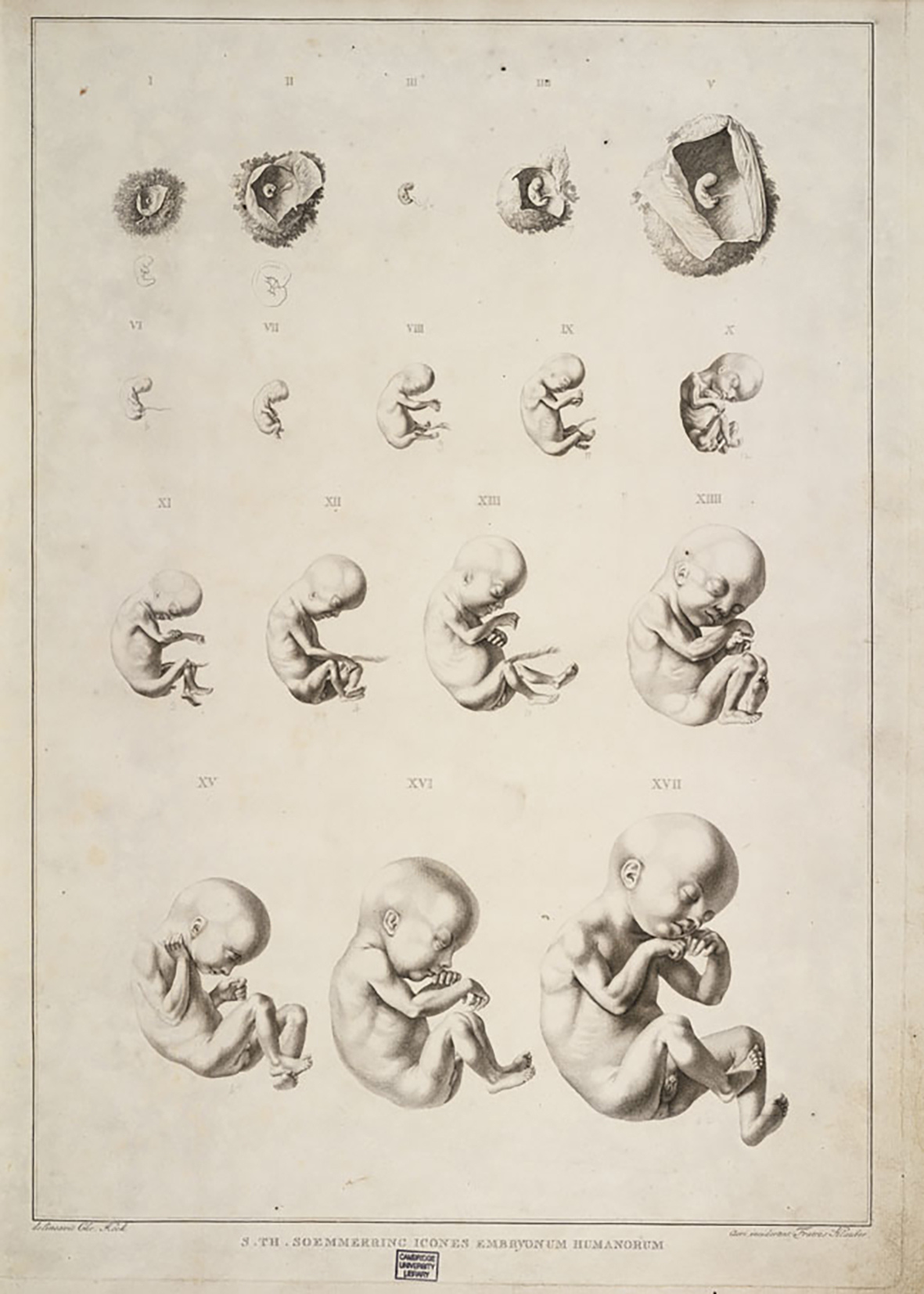
From his collection, Soemmerring chose those fetuses which recommended themselves to him as types due to their “beauty,” corresponding with their age. Already in the third week, he “sees'' in the egg, which he has placed in diluted wine, a three-week-old body with bulbous extensions, hardly longer than a “Parisian line.” The genitals sprout, (efflorescunt) early in the second month. The penis is all the more prominent the younger the embryo is, and resembles a peeled glans. The virginalis (maiden part) can occasionally be recognized as a small cleft in the second month. In the third month the clitoris almost resembles the erect male member, so that when viewed from the side, the female embryo could be confused with the male embryo. Soemmerring refers to his “Description of Miscarriages” (1791), in which he found that female fetuses are more likely to be lost, a proportion in premature birth that is also true in de embryonibus monstrosis.
The non-genital sexual characteristics, he says, are much more visible than the genitals. But nowhere has he read of this 'significant' sex difference in fetuses from other authors, although these features are scarcely absent in any embryo, however small and rotten, unless the object is clearly a monster. The most significant difference lies in the structure of the thorax, which in the female embryo becomes narrower like an amphora and its shoulders correspondingly more sloping. Not only does the abdomen begin higher up, but it is so curved that the genitals are like little sacks; if one dares to speak metaphorically - according to Soemmerring - they look like a tumor (tumidulum diceres). The gender can also be identified by the shape of the head, the strength of the hands, the forearms and the heels. Here the anatomist is speaking a few years before Goethe coined the word “morphology.” The embryos give him a welcome opportunity to examine the characteristics of the fair sex, produced by the dissector, in their prenatal destiny. I know the type of this “desired seeing” from earlier anatomists, but it was not then a question of sex difference, whether the beak-mouthed children observed by Kerckring or the female eggs of Govrard Bidloo that were documented as having been seen. Despite his readings of Kant, Soemmerring still inspires the anatomical libido videndi, the visual curiosity from the Theatrum Anatomicum.
Iconological interpretation
I arrive at completely different insights if I do not concern myself with the explicit motives of the inquiring gaze at the penis or heels, and if I do not deal with the poetic legends accompanying the tablets, but rather pursue the question of how Soemmerring himself wanted to shape the process of scientific illustration. We know his opinion about this, because he sums it up succinctly in the introduction to the Icones.
First, Soemmerring wonders why the embryonic form has not been “seen” so far. What prejudices prevented even anatomists from wanting to recognize the embryo as a coming child? According to Soemmerring, “God knows what old wives' tales mislead not only lay people but also artists who consider the form of the human embryo to be repulsive, even unbearable or monstrous.”18) That is the first reason why “what was hidden in the womb” remained excluded from the anatomical representation. Soemmerring names the old traditional reluctance to perceive the forma substantialis of man in pre-childhood stages. He takes note of the willingness to expect anything and everything surprising from the womb, and to take it for a monster if it does not look like a child.
Finally, he names a fourth reason that had previously made it difficult to see: an intentional looking away. What midwives brought to doctors, what was kept in cabinets of curiosities and anatomy rooms, and what had faded in spirits of wine, could hardly arouse admiration: “They don't want to see (intueri) what corresponds to the order of nature, but what corresponds to their opinion: so they despise not only the rotten and spoiled fruit they lay their hands on, but even those most perfect for their stage.” Soemmerring argues for the possibility that a being can appear morphologically different at different moments in its development and yet appear in all its beauty.
The choice of specimens is thus guided by the perception of their “beauty.” The anatomist deliberately restrains the draftsman's attention: the position of the fetal limbs is left “as the homunculi were left to me.”19) But in no way are the wrinkles, swellings, and disfigurements, which result from the storage in Schnaps, slavishly reproduced or made to look ridiculous20): only what the anatomist recognizes as “significant” should be made visible by the draftsman. Soemmerring wants effigies, that is, a portrait, only not at the expense of fine details. But an archetypus should also be depicted: “I placed them all in such a way that the light falls on them at an angle of 40 degrees, and my draftsman was primarily confronted by the head, forehead, nose, cheeks, mouth, the shape of the chest and the genitals.”21) With these precautions, Soemmerring wants a paradoxical combination: the accuracy of a unique portrait and the depiction of a type.
In order to realize this paradoxical combination of individuality and type, he must, three decades before the very first photograph by Daguèrre, shut the eye in the production of scientific imaging. He breaks with the anatomical Renaissance tradition, which wanted to convey an image of the body according to the rules of central perspective, giving the viewer's eye the same impression as if the objectum were lying in front of him. Inspired by an exchange of views between his older friend Petrus Camper and the Leiden anatomist Bernhard Siegfried Albinus, Soemmerring regards “perspective” as the form in which illusion is brought into the picture. The draftsman Christian Koeck, whom he trained for years, is not supposed to reproduce what his eye sees, but he is supposed to create an outline of the fetus after architectural-geometric surveying (more geometrico). He does not want to represent the object as he or the draftsman sees it, but rather he wants to have the object measured and the measurement results given graphic expression. The draftsman must locate each point of the object through a double grid, aiming at each detail and drawing it as if seen at right angles from a great distance. This allows the object to be represented as it were, in itself, since it is no longer related to the viewer's eye in perspective.
Soemmerring wants a simulacrum des objectes and not a facsimile of what the eye sees. He does not want an image, but a construct. He knows that the figures he has had engraved can never be seen that way with the naked eye. The simulacrum creates a new kind of objectivity: a forcibly distanced view of objects and also of their relationships to each other.22)
Soemmerring thoroughly robs the sensual act of seeing of its innocence and demands the contemplation of a constructed “reality”. He agrees with Peter Camper that central perspective serves to carry the distortion inherent in sensory perception into the picture. To exclude the sensual and therefore the deceptive perception of the observer, he does not want perspective but isometric projection, i.e., a reconstructed “reality”. His pre-childhood human series from 1799 only seems understandable to me because of this desire for a viewpoint without a point-of-view.
The metamorphosis of the unborn child into a “fetus” begins with the planimetric-architectural (planimetrisch) drawing method used by Soemmerring. From that moment on, the depiction of the fetus remains technogenic, as far as I have been able to trace into the nineteenth and twentieth centuries. The visual concept of the unborn as a “pre-child” being can only be understood, from Koeck's drawings, later microphotography, and X-ray fluoroscopy to ultrasound, if one understands it as a history of the interpretation of mechanically produced records.
History of gender relations as the basis for a historical understanding of woman today
I began with a history of the representation of the unborn, an iconographic history which has already been studied by many authors. As we have seen, the body of the unborn was often a surface onto which researchers projected ideas about the special position and competence that nature had assigned to women.
I unreservedly accept that the history of the scientific production of concepts and ideas about the female body has been a basis for the legal, social, economic, and cultural position of women since the later eighteenth century. It has also progressively marked women's self-image and experience. The scientific characterization of female “biology” is and remains a basis for the historical investigation of group identity and of the formation of interests, as Karin Hausen puts it. This remained so until the late 19th century.23)
I would like to complement, or add a layer to, this research into the historical genesis of women's gender perception, based on experienced discrimination, however necessary it may be.
I am trying to engage with that disembodiment of perception and that sentimental understanding of scientific fact that Soemmerring's Icones gave me the opportunity to discuss. Today, in my opinion, the historicity of a tangible, sensual difference between woman and man is threatened by their abstract reduction to two historical agents. This difference is increasingly being formulated in a deconstructivist fashion, so that it is disguised or explicitly merged into a system-theoretical binomial.
I am of the opinion that around 1800 a polarization takes place in the sensual perception of the sexes that, paradoxically, begins to produce an abstract leveling of what had formerly been understood to be their difference. I consider it the central theme of gender history to follow the consequences of this process in the various areas of society up to the present. The type of “gender” that emerges is modern. The divergence between the experience of quickening by which a woman once knew her pregnancy, and what contemporary people take for granted about “sexuality” and the being of the fetus, has a history.